Figure 1
A Rare case of synchronous primary malignancies of gall bladder and ovary
Ridhi Narang*, T Das, M Dagar, M Srivastava, S Bhalla and I Ganguli
Published: 06 September, 2018 | Volume 1 - Issue 2 | Pages: 052-055

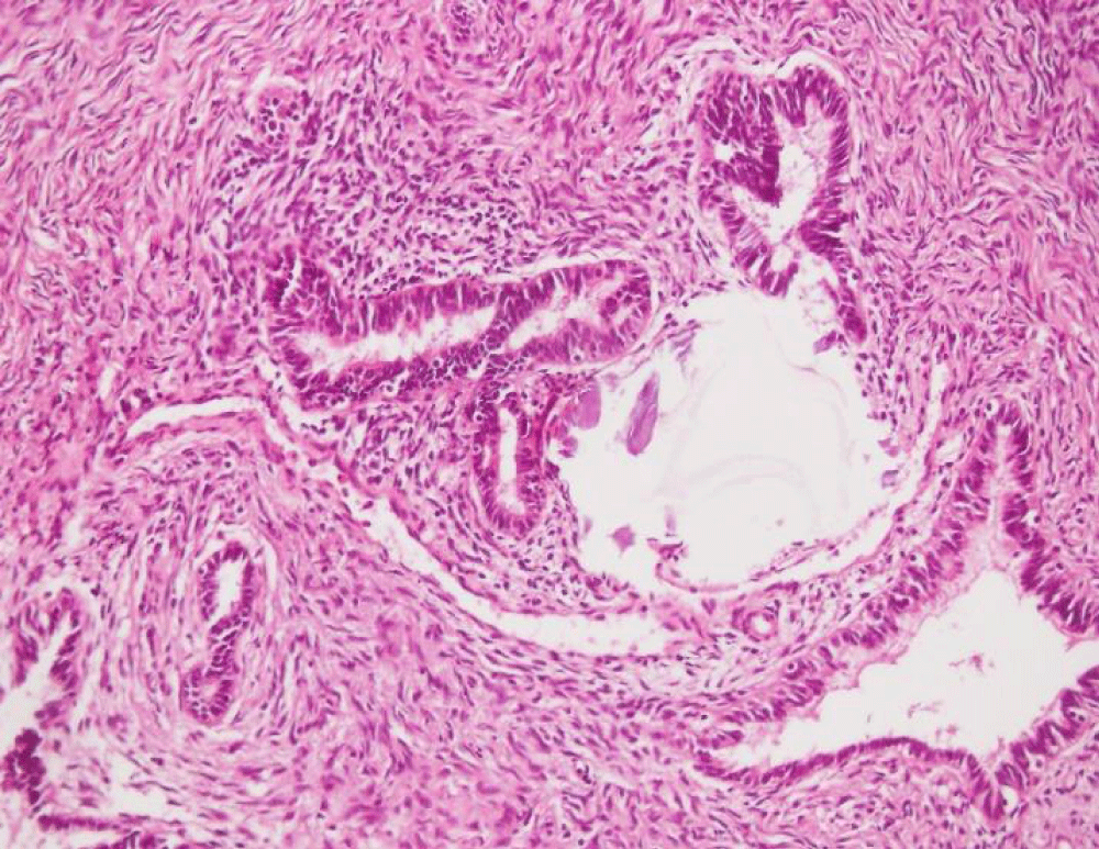
Figure 1:
Section from the ovarian tumor shows cystic tumor lined by complex branching papillae, the papillae wall lined by multiple layers of tumor cells showing hyper chromatic nuclei, nuclear pleomorphism and moderate eosinophilic cytoplasm.
Read Full Article HTML DOI: 10.29328/journal.cjog.1001008 Cite this Article Read Full Article PDF
More Images
Similar Articles
-
TMD and pregnancy?Afa Bayramova*. TMD and pregnancy?. . 2018 doi: 10.29328/journal.cjog.1001001; 1: 001-006
-
Is It Possible to End Female Circumcision in Africa?Fiona Dunn*. Is It Possible to End Female Circumcision in Africa? . . 2018 doi: 10.29328/journal.cjog.1001002; 1: 007-013
-
Screening of Gestational diabetes mellitusGehan Farid*,Sarah Rabie Ali*,Reem Mohammed Kamal. Screening of Gestational diabetes mellitus . . 2018 doi: 10.29328/journal.cjog.1001003; 1: 014-023
-
The Case of the Phantom Trophoblastic TumorBenedict B Benigno*. The Case of the Phantom Trophoblastic Tumor. . 2018 doi: 10.29328/journal.cjog.1001004; 1: 024-025
-
Maternal and fetal outcome of comparative study between old & adopted new value of screening of Gestational Diabetes Mellitus in tertiary centre in Saudi ArabiaGehan Farid*,Reem Mohammed Kamal*,Mohamed AH Swaraldahab,Sarah Rabie Ali. Maternal and fetal outcome of comparative study between old & adopted new value of screening of Gestational Diabetes Mellitus in tertiary centre in Saudi Arabia. . 2018 doi: 10.29328/journal.cjog.1001005; 1: 026-034
-
Small cell carcinoma of the ovary with hypercalcemia: Case report and review of the literatureGerday A*,Marbaix E,Squifflet JL,Mazzeo F,Luyckx M. Small cell carcinoma of the ovary with hypercalcemia: Case report and review of the literature. . 2018 doi: 10.29328/journal.cjog.1001006; 1: 035-044
-
Perinatal Morbidity & Mortality following repeat Cesarean section due to five or more previous Cesarean Section done in Tertiary centre in KSASomia Osman,Gehan Farid*,Reem Mohamed Kamal,Sarah Rabie Ali,Mohamed AH Swaraldahab. Perinatal Morbidity & Mortality following repeat Cesarean section due to five or more previous Cesarean Section done in Tertiary centre in KSA. . 2018 doi: 10.29328/journal.cjog.1001007; 1: 045-051
-
A Rare case of synchronous primary malignancies of gall bladder and ovaryRidhi Narang*,T Das,M Dagar,M Srivastava,S Bhalla,I Ganguli. A Rare case of synchronous primary malignancies of gall bladder and ovary. . 2018 doi: 10.29328/journal.cjog.1001008; 1: 052-055
-
Effectiveness of the lifestyle modifications in prevention and control of sexually transmitted diseases (STDs): Focus on Islamic lifestyleMohammad Rabbani Khorasgani*. Effectiveness of the lifestyle modifications in prevention and control of sexually transmitted diseases (STDs): Focus on Islamic lifestyle. . 2018 doi: 10.29328/journal.cjog.1001009; 1: 056-057
-
Septic arthritis of left shoulder in pregnancy following minor hand injuryNeelam Agrawal,Rhoughton Clemmey,Shamma Al-Inizi*. Septic arthritis of left shoulder in pregnancy following minor hand injury. . 2018 doi: 10.29328/journal.cjog.1001010; 1: 058-060
Recently Viewed
-
Drug Rehabilitation Centre-based Survey on Drug Dependence in District Shimla Himachal PradeshKanishka Saini,Palak Sharma,Bhawna Sharma*,Atul Kumar Dubey,Muskan Bhatnoo,Prajkta Thakur,Vanshika Chandel,Ritika Sinha. Drug Rehabilitation Centre-based Survey on Drug Dependence in District Shimla Himachal Pradesh. J Addict Ther Res. 2025: doi: 10.29328/journal.jatr.1001032; 9: 001-006
-
Causal Link between Human Blood Metabolites and Asthma: An Investigation Using Mendelian RandomizationYong-Qing Zhu, Xiao-Yan Meng, Jing-Hua Yang*. Causal Link between Human Blood Metabolites and Asthma: An Investigation Using Mendelian Randomization. Arch Asthma Allergy Immunol. 2023: doi: 10.29328/journal.aaai.1001032; 7: 012-022
-
Impact of Latex Sensitization on Asthma and Rhinitis Progression: A Study at Abidjan-Cocody University Hospital - Côte d’Ivoire (Progression of Asthma and Rhinitis related to Latex Sensitization)Dasse Sery Romuald*, KL Siransy, N Koffi, RO Yeboah, EK Nguessan, HA Adou, VP Goran-Kouacou, AU Assi, JY Seri, S Moussa, D Oura, CL Memel, H Koya, E Atoukoula. Impact of Latex Sensitization on Asthma and Rhinitis Progression: A Study at Abidjan-Cocody University Hospital - Côte d’Ivoire (Progression of Asthma and Rhinitis related to Latex Sensitization). Arch Asthma Allergy Immunol. 2024: doi: 10.29328/journal.aaai.1001035; 8: 007-012
-
The Color of Diseases and Herbs-chromatic Illustrating the Yin-yang Regulation Theory in Traditional Chinese MedicineHuigang Liu*. The Color of Diseases and Herbs-chromatic Illustrating the Yin-yang Regulation Theory in Traditional Chinese Medicine. Ann Biomed Sci Eng. 2025: doi: 10.29328/journal.abse.1001034; 9: 001-004
-
Exploring the Potential of Medicinal Plants in Bone Marrow Regeneration and Hematopoietic Stem Cell TherapyUgwu Okechukwu Paul-Chima*,Alum Esther Ugo. Exploring the Potential of Medicinal Plants in Bone Marrow Regeneration and Hematopoietic Stem Cell Therapy. Int J Bone Marrow Res. 2025: doi: 10.29328/journal.ijbmr.1001019; 8: 001-005
Most Viewed
-
Feasibility study of magnetic sensing for detecting single-neuron action potentialsDenis Tonini,Kai Wu,Renata Saha,Jian-Ping Wang*. Feasibility study of magnetic sensing for detecting single-neuron action potentials. Ann Biomed Sci Eng. 2022 doi: 10.29328/journal.abse.1001018; 6: 019-029
-
Evaluation of In vitro and Ex vivo Models for Studying the Effectiveness of Vaginal Drug Systems in Controlling Microbe Infections: A Systematic ReviewMohammad Hossein Karami*, Majid Abdouss*, Mandana Karami. Evaluation of In vitro and Ex vivo Models for Studying the Effectiveness of Vaginal Drug Systems in Controlling Microbe Infections: A Systematic Review. Clin J Obstet Gynecol. 2023 doi: 10.29328/journal.cjog.1001151; 6: 201-215
-
Causal Link between Human Blood Metabolites and Asthma: An Investigation Using Mendelian RandomizationYong-Qing Zhu, Xiao-Yan Meng, Jing-Hua Yang*. Causal Link between Human Blood Metabolites and Asthma: An Investigation Using Mendelian Randomization. Arch Asthma Allergy Immunol. 2023 doi: 10.29328/journal.aaai.1001032; 7: 012-022
-
An algorithm to safely manage oral food challenge in an office-based setting for children with multiple food allergiesNathalie Cottel,Aïcha Dieme,Véronique Orcel,Yannick Chantran,Mélisande Bourgoin-Heck,Jocelyne Just. An algorithm to safely manage oral food challenge in an office-based setting for children with multiple food allergies. Arch Asthma Allergy Immunol. 2021 doi: 10.29328/journal.aaai.1001027; 5: 030-037
-
Impact of Latex Sensitization on Asthma and Rhinitis Progression: A Study at Abidjan-Cocody University Hospital - Côte d’Ivoire (Progression of Asthma and Rhinitis related to Latex Sensitization)Dasse Sery Romuald*, KL Siransy, N Koffi, RO Yeboah, EK Nguessan, HA Adou, VP Goran-Kouacou, AU Assi, JY Seri, S Moussa, D Oura, CL Memel, H Koya, E Atoukoula. Impact of Latex Sensitization on Asthma and Rhinitis Progression: A Study at Abidjan-Cocody University Hospital - Côte d’Ivoire (Progression of Asthma and Rhinitis related to Latex Sensitization). Arch Asthma Allergy Immunol. 2024 doi: 10.29328/journal.aaai.1001035; 8: 007-012

If you are already a member of our network and need to keep track of any developments regarding a question you have already submitted, click "take me to my Query."


